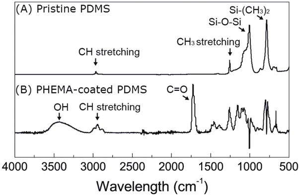
Fig. 2. Qualitative spectroscopic analysis of uncoated and PHEMA-coated PDMS ventricular catheters.
ATR-FTIR spectra of (A) pristine and (B) iCVD PHEMA-coated PDMS catheters. The PDMS background was subtracted out of the PHEMA-coated PDMS spectrum. The OH (3434 cm−1) and C=O (1726 cm−1) peaks of PHEMA suggest that PHEMA was successfully deposited on the catheters.

