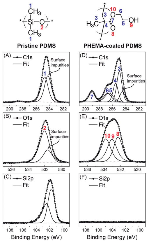
Fig. 3. Quantitative spectroscopic analysis of uncoated and PHEMA-coated PDMS ventricular catheters.
High resolution C1s, O1s, and Si2p XPS spectra of pristine (A–C) and iCVD PHEMA-coated (D–F) PDMS catheters with their corresponding fitted peaks and peak assignments (see Table 1 also). The C:O:Si ratio of pristine PDMS, estimated at 2.1:1.0:1.0, agrees well with stoichiometric PDMS. The C:O ratio of the PHEMA coating, estimated at 2.5:1.0, is close to that of stoichiometric PHEMA. The disappearance of Si in the PHEMA-coated sample indicates the PHEMA coating is sufficiently thick to prevent the underlying PDMS substrate from being probed.

