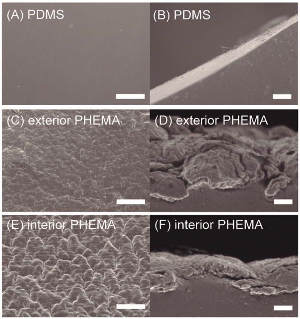
Fig. 4. Surface morphological analysis of uncoated and PHEMA-coated ventricular catheters.
Top- and side-view SEM images of pristine (A,B) and iCVD PHEMA-coated (C,D and E,F) PDMS catheters. The latter pairs of images are for PHEMA coatings on the exterior (C,D) and interior (E,F) catheter walls, respectively. Surface roughness and feature sizes are related to the nucleation density for polymer growth that is affected by the accessibility of activated initiator species. Scale bar is 10 and 1 μm for top and side views, respectively.

