
Figure 1.
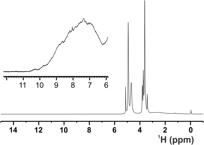
1D 1H NOESY NMR spectrum (mixing 20 ms) acquired at the solid-state on the ANSII-GNPs sample using a spectrometer operating at 800 MHz, 1H Larmor frequency, at ~282 K and MAS of 60 kHz. The sharp signals of the methylene group of the PEG chains and of the water are visible around 3.6 and 4.9 ppm, respectively. The top panel displays an enlargement, with a different intensity scale, of the region between 6 and 12 ppm, where the protein amide proton resonates. The high spreading of these signals is a positive marker of the protein folding.