Figure 6.
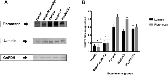
Analysis of FN and LN proteins expression of Balb/c mice bearing 4T1 breast tumor. The animals were treated with Magh-cit or Rh2(H2cit)4 or Magh-Rh2(H2cit)4. (A) western blotting analysis shows a band of approximately 250 kDa (FN) and 225 kDa (LN) in the total protein extracts from normal and tumor tissues from mice. (B) Quantification of relative protein expression of LN and FN found in western blotting analysis. GAPDH was used as internal control. The healthy and Magh-Rh2(H2cit)4 treated groups showed markedly statistically significant reduction when compared to the control group. The values shown are the mean ± SEM *p < 0.05 and analyzed by an ANOVA; Tukey’s post hoc test.

