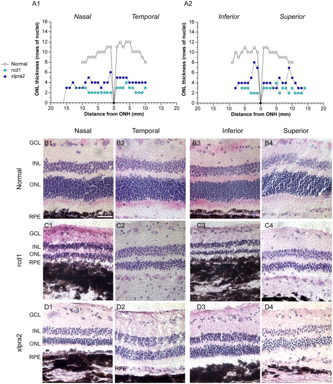
Figure 1.
ONL count in normal and mutant retinas. (A1–2) Biaxial spidergraphs of outer nuclear layer thickness in the nasal-temporal and superior-inferior meridians for normal, rcd1 and xlpra2 retinas. H&E stained photomicrographs illustrating ONL thickness differences in the nasal, temporal, superior and inferior meridians of retina in a (B1–4) normal dog, (C1–4) rcd1 dog, and (D1–4) xlpra2 dog at 3000 ± 500 microns from the optic nerve head. RPE: Retinal Pigment Epithelium; ONL: Outer Nuclear Layer; INL: Inner Nuclear Layer; GCL: Ganglion Cell Layer; ONH: Optic Nerve Head.

