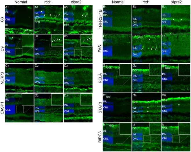
Figure 5.
Immunohistochemical localization of select pathway proteins in normal and mutant retina. (A1–3) Complement component C3; (B1–3) Complement component and component of the membrane attack complex (MAC) C9; (C1–3) Inflammasome component NLRP3; (D1–3) Apoptosis executor and IL1β activator protein Caspase-1 (CASP1); (E1–3) TNF superfamily receptor TNFRSF1B; (F1–3) Cell surface death receptor FAS; (G1–3) Transcription factor of NFκB family, RELA (p65); (H1–3) Transcription activator protein of STAT family, STAT3; (I1–3) Anti-apoptotic protein BIRC3. RPE: Retinal Pigment Epithelium; ONL: Outer Nuclear Layer; OPL: Outer Plexiform Layer; INL: Inner Nuclear Layer; GCL: Ganglion Cell Layer.

