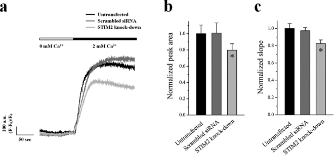
Figure 4.
Decreased SOCE by STIM2-knockdown. The Ca2+ in the SR of the STIM2-knockdown myotubes was depleted by the treatment of TG (2.5 μ M) in the absence of extracellular Ca2+. Extracellular Ca2+ (2 mM) was applied to the myotubes to induce SOCE. (a) A representative trace for each group is shown. The results are summarized as bar graphs for the area under the peaks (b) or the slope at the rising phase of the peaks (c). *Significant difference compared with the untransfected control (P < 0.05). The values are presented as the mean ± S.E. for the number of myotubes shown in the parentheses of Table 4. SOCE was significantly decreased by STIM2-knockdown.

