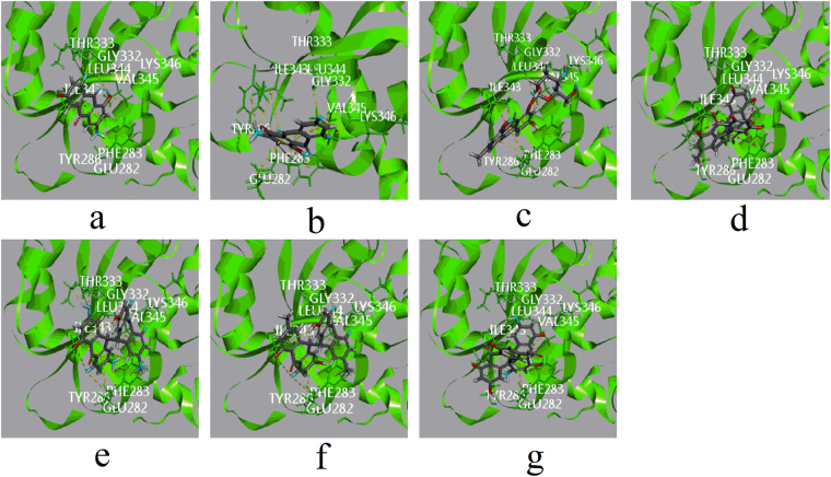
Figure 4.
Computational docking of ligands in site F. The interaction between ligands and amino acid residues of UGT1A1 (a) emodin with ILE343, VAL345, LEU344, THR333 and GLY332. (b) Citreorosein with ILE343, VAL345, PHE283, LYS346 and GLY332. (c) Emodin-8-O-glc with ILE343, VAL345, PHE283, LYS346, GLY332 and LEU344. (d) Trans-emodin dianthrone (10αH/10βH) with VAL345, ILE343 and LY332. (e) Trans-emodin dianthrone (10 βH/10αH) with VAL345, ILE343, GLY332, THR333, LYS346 and VAL345 (f) cis-emodin dianthrone (10αH/10αH) with VAL345, ILE343, GLY332, LYS346 and VAL345 (g) cis-emodin dianthrone (10βH/10βH) with PHE283, VAL345, ILE343 and GLY332). All involved ligands and side chains by element had been coloured.

