
Figure 1.
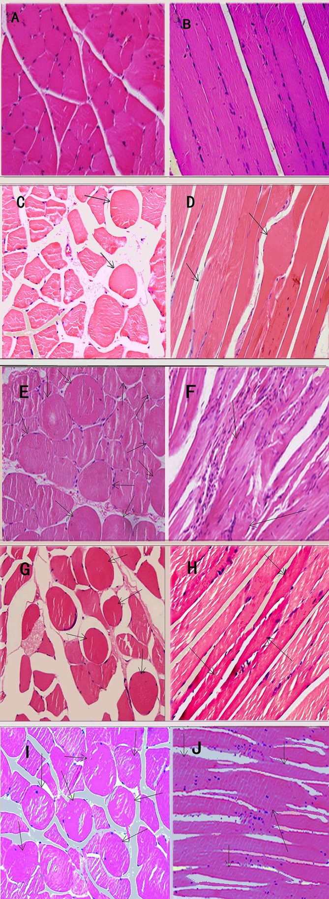
Light microscope views of muscle fibres with haematoxylin and eosin staining, ×400. In control group 1 (CG1), the muscle cells showed polygonal shapes of uniform size (A). Moreover, tight and ordered arrangement was found in the longitudinal section (B). In control group 2 (CG2), very few large round muscle cells could be seen in cross-section (C) except that a long thin fibre was observed connecting to a knot (D). In the 4-week (4W), 8W and 12W groups, several large and small hyperchromatic rounded muscle cells appeared (black arrows) in cross-section (E, G, I) and several fusiform muscle fibres (arrows) connecting thinning fibres matched with rounded muscle cells (F, H, J). In the 8W and 12W groups, continuous beaded muscular fibres with enlargement in the middle (black arrow) and attenuation at both ends and infiltration of slight inflammatory cells could be observed in longitudinal section (H and J). In both sections, spaces between fibres became the width in the 8W and 12W groups.