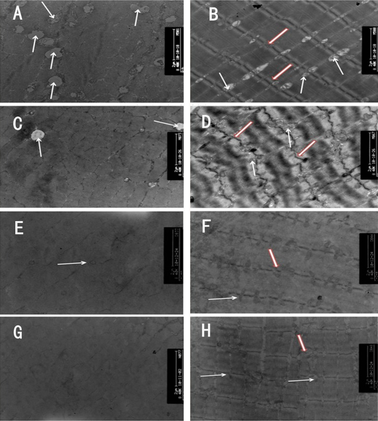
Figure 2.
Transmission electron microscope views of muscle fibres, ×8400. In control group 1 (CG1), large numbers of ovate mitochondria (white arrows) with ridge-like structures inside and intact myofilaments in well-arranged and clean-cut sarcomeres were observed either in cross-section or in longitudinal section (A, B). In the 4-week (4W), 8W and 12W groups, the number of mitochondria (white arrows) was reduced or even disappeared in cross-section. Their shapes were changed into a circle, and their ridge-like structures decreased or disappeared (C, E and G). The arrangement of sarcomeres was abnormal, and the myofilaments were disarranged and blurred in longitudinal section (D, F and H). The Z-line (hollow red arrow) became thinner than that in CG1 (D, F and H), around which fewer mitochondria were found. Moreover, the Z-line in the 4W rooked like a drifting wave-like line (D).

