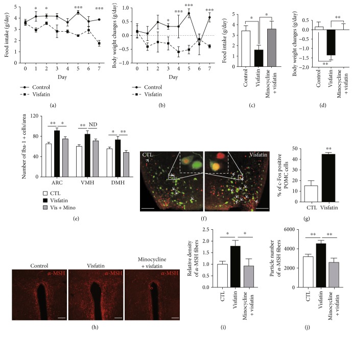
Figure 4.
Visfatin leads to anorexia and body weight loss by controlling microglia and POMC neuronal axis. (a) Food intake and (b) body weight change were measured for seven days, after daily icv administration of visfatin (n = 5 mice/group). Visfatin-induced anorexia (c) and body weight loss (d) were significantly reversed by administration of minocycline (Mino) for 3 consecutive days prior to icv visfatin treatment (n = 4 mice/group). (e) The increased number of microglial cells induced by icv injection of visfatin was effectively rescued by preadministration of minocycline (n = 8 sections/4 mice for control; n = 8 sections/4 mice for visfatin; n = 8 sections/4 mice for minocycline + visfatin). (f) Representative images and (g) quantitative analysis of the number of c-Fos-positive POMC cells following icv injection of visfatin (n = 8 sections/4 mice for control (CTL); n = 8 sections/4 mice for visfatin). (h) Representative images show α-MSH immunosignals in the PVN. Visfatin-induced increase in (i) relative density and (j) particle number of α-MSH fiber signals was completely reversed by pretreatment of minocycline (n = 8 sections/4 mice for control; n = 8 sections/4 mice for visfatin; n = 8 sections/4 mice for minocycline + visfatin). White arrows: double-labeled c-Fos and POMC positive cells. Scale bar = 100 μm. Results are presented as mean ± SEM. ∗P < 0.05, ∗∗P < 0.01, and ∗∗∗P < 0.005.

