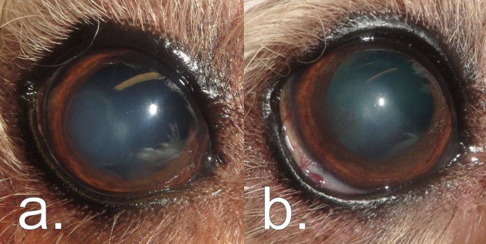
FIG 1:
Thelazia callipaeda-associated pathology in the right eye of case 3 showing (a) superficial ventrolateral corneal ulceration at the initial point of referral, and (b) re-epithelialisation associated with ventral bulbar conjunctival hyperaemia, ventrolateral corneal oedema and ventrolateral superficial corneal vascularisation 21 days after flushing and removal of a single male T callipaeda specimen from the ventral conjunctival fornix.

