FIG 2:

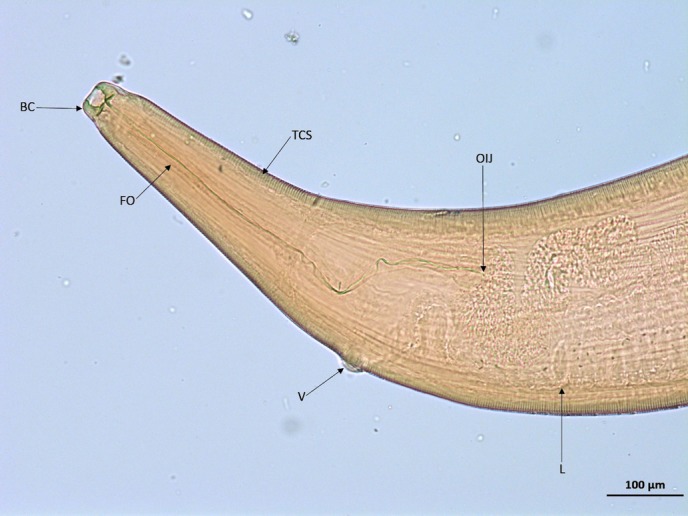
Light micrograph of female Thelazia callipaeda, anterior. BC, buccal capsule; FO, filariform oesophagus; L, L1 larvae; OIJ, oesophagus–intestinal junction; TCS, transverse cuticular striations; V, vulva (×10).

Light micrograph of female Thelazia callipaeda, anterior. BC, buccal capsule; FO, filariform oesophagus; L, L1 larvae; OIJ, oesophagus–intestinal junction; TCS, transverse cuticular striations; V, vulva (×10).