
Figure 5. Fibrous capsule measurements as seen in Case 10L implanted with straight-banded electrode of Nucleus 22 device.
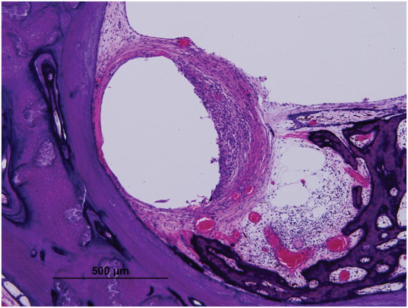
Low (A) and high (B) magnification view at LB (large arrow).
Low (C) and high (D) magnification view at UB (large arrow). Asterisk indicates the electrode track; Lines indicate the thickness of fibrous tissue at each point: S, superior; M, medial; I, inferior; SV, scala vestibuli; ST, scala tympani; }, new bone formation; small arrow, osseous spiral lamina; IAC, internal acoustic canal