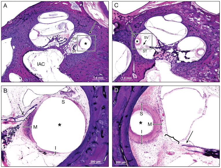
Figure 7. Intracochlear damage and fibrous capsule thickness.
An asymmetric fibrous tissue thickness in Case 2L (Low (A) and high (B) magnification view at lower basal turn of the cochlea), which was implanted with an AB perimodiolar electrode resulted in fracture of the osseous spiral lamina (arrow in Figure B) and marked displacement of the basilar membrane.
A symmetric fibrous tissue thickness in Case 10L (Low (C) and high (D) magnification view at lower basal turn of the cochlea), which was implanted with a Nucleus 22 straight electrode and resulted in marked displacement of the basilar membrane (}), but with an intact osseous spiral lamina (arrow).
Large arrow marks basal turn of cochlea; asterisk (*) indicates the electrode track; S, superior; M, medial; I, inferior: represent the thickness of fibrous tissue in each point; SV, scala vestibuli; ST, scala tympani; IAC, internal acoustic canal.

