
Figure 3.
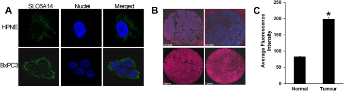
SLC6A14 protein expression is up‐regulated in pancreatic cancer. (A) Immunofluorescence analysis of SLC6A14 protein in HPNE and BxPC‐3 cells (green). Nuclei were stained with Hoechst (blue). (B) Representative images of SLC6A14 protein expression in two normal and two tumour pancreatic tissues from a TMA. Images are shown at 20× magnification. (C) Histogram showing SLC6A14 staining intensity in normal and tumour pancreatic tissues assessed from the whole TMA (normal tissue, n = 5; tumour tissue, n = 53). Data are given as means ± SEM. *P < 0.05.