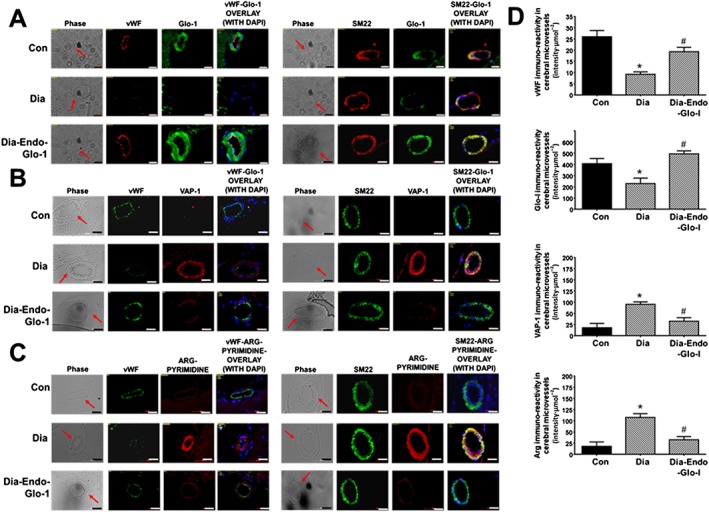
Figure 8.

Panels A, B and C show representative immunohistochemical data for vWF, SM22‐α, DAPI, Glo‐I, VAP‐I and Arg in cortical microvessels in 20‐μm thick brain slices (Bregma −3.0 to −3.3) from Con, Dia and Dia‐Endo‐Glo‐I animals. White bar at bottom of each image = 20 μm. Graphs on the right (Panels D), starting from the top, show quantification of the amount of vWF, Glo‐I, VAP‐I and Arg in cortical arterioles in brains from Con, Dia and Dia‐Endo‐Glo‐I animals. Values shown are means ± SEM for 40 vessels from n = 5–6 rats. * P < 0.05, significantly different from Con; # P < 0.05, significantly different from Dia.
