Figure 5.

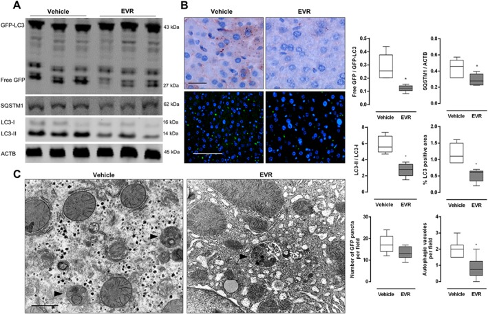
Continuous long‐term (28 days) everolimus treatment results in a decrease in autophagic markers in vivo. GFP‐LC3 mice received osmotic minipumps releasing either everolimus (EVR, 1.5 mg kg−1 day−1) or vehicle for a period of 28 days. Prior to killing, mice received an i.p. injection of 100 mg kg−1 chloroquine. (A) Western blotting was performed to assess the ratio of free GFP/GFP‐LC3 and LC3‐II/LC3‐I as well as the accumulation of SQSTM1. (B) Liver samples were immunohistochemically stained for LC3 (upper panels) or analysed with fluorescence microscopy for GFP positive puncta (lower panels). Scale bar = 100 μm. (C) Autophagic vacuoles (arrow heads) were detected in ultrathin sections using TEM. Scale bar = 1 μm (n = 6 for both groups, *P < 0.05, significantly different from vehicle).
