
Fig. 1.
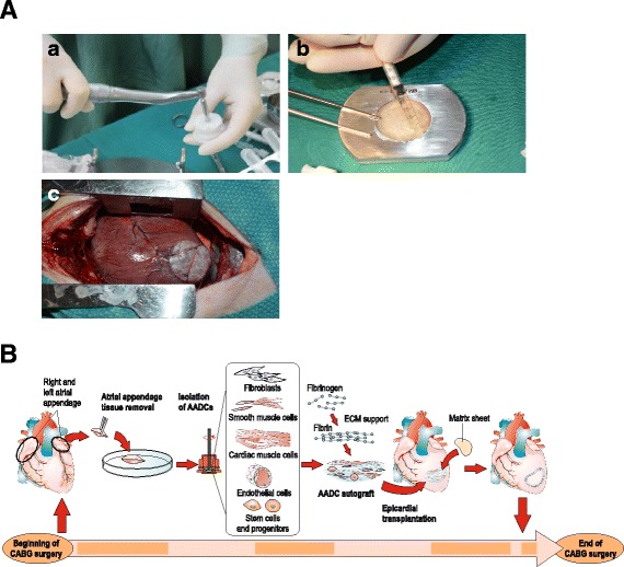
a Preparing the AADC-sheet. (A) The atrial appendage tissue is processed with cell therapy tissue homogenizer (Rigenera-system). (B) The micrografts are secured to extracellular matrix sheet (Cormatrix®) by using a fibrin sealant (Tisseel™). (C) The AADC sheet is placed to the myocardium in the location of infarction scar (animal model). b Administration of therapy during CABG surgery. Figure reproduced from our article by Lampinen et al. (Current Gene Therapy, 2015)