Figure 2.

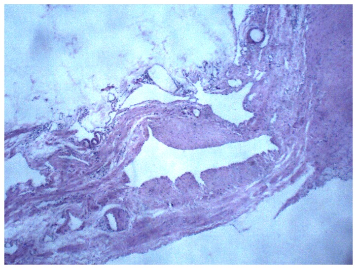
Histopathological section of the hemolymphangioma from case 1 with hematoxylin and eosin staining highlighting the blood and lymphatic vessels. Magnification, ×100.

Histopathological section of the hemolymphangioma from case 1 with hematoxylin and eosin staining highlighting the blood and lymphatic vessels. Magnification, ×100.