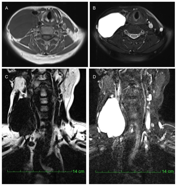
Figure 4.
Magnetic resonance imaging results from case 2. A cystic mass was observed on the right side of the neck with (A) hypo-intensity on T1-weighted images (B) and hyper-intensity on T2-weighted images. The tumor demonstrated (C) hypo-intensity on the water-suppression images and (D) significant hyper-intensity on the fat-suppression images.

