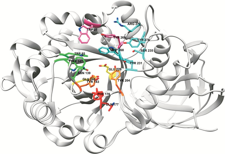
Fig. (6).
A typical active site of a GH30_8 xylanase with glucuronoxylanase activity, here illustrated by BcXyn30C from Bacillus subtilis, PDB 3KLO [104]. The amino acids are colored in accordance with subsite-numbering; -2: pink, -2 substitution: cyan, -1: green, +1: orange and +2: red. The catalytic residues are colored in yellow. Tyr204 is also a part of subsite +1 (orange), Trp268 is also a part of subsite -1 (green) and Arg272 as well as Trp27 are possibly part of subsite -3. (The color version of the figure is available in the electronic copy of the article).

