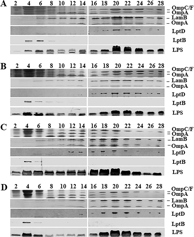
FIG 5.
Membrane fractionation of wild-type, lptA41, and suppressor strains. Cultures of PS001 (lptA+) (A), PS003 (lptA41) (B), PS102 (mlaA102) (C), and PS103 (lptA42 opgH103) (D) strains were grown to an OD600 of ∼0.6. Crude extracts were fractionated on a sucrose density gradient. Fractions were collected from the top of the gradient. The profiles of the major OM porins (OmpC/F-OmpA) were determined by SDS-PAGE, followed by Coomassie blue staining. LamB-OmpA and LptD profiles were determined as OM markers by immunoblotting using anti-LamB and anti-LptD antibodies, respectively. LptB was detected as an IM marker using anti-LptB antibody. LPS distribution across fractions was determined by Tricine SDS-PAGE and immunoblotting using anti-LPS WN1 222-5 monoclonal antibody. The numbers are fraction numbers.

