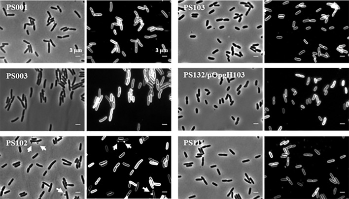
FIG 7.
Morphology of lptA41 and suppressor cells. Shown are phase-contrast and membrane stain (FM5-95) images of PS001 (lptA+), PS003 (lptA41), suppressor strains PS102 (mlaA102) and PS103 (lptA42 opgH103), PS132/pOpgH103 (ΔopgH ectopically expressing OpgH103 mutant protein), and PS111 (lptA42). Cell length measurements are reported in Table 5. The arrows in suppressor strain PS102 indicate OMVs.

