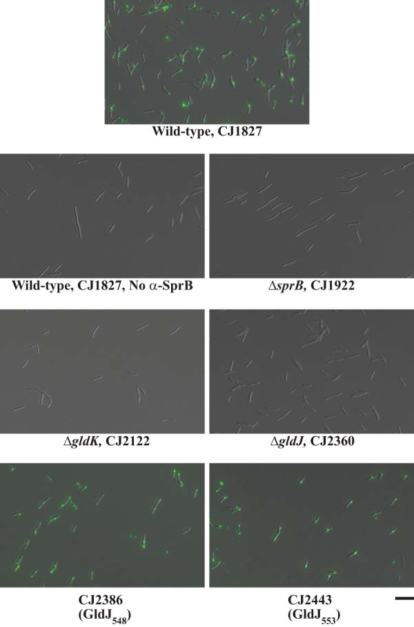
FIG 11.

Detection of surface-localized SprB by immunofluorescence microscopy. Cells of wild-type and mutant F. johnsoniae were exposed to anti-SprB antiserum followed by F(ab′) fragment of goat anti-rabbit IgG conjugated to Alexa Fluor 488. Cells of mutants CJ2386 and CJ2443 produce truncated versions of GldJ (548 and 553 aa, respectively). Images were recorded by immunofluorescence microscopy and by differential interference contrast (DIC) microscopy and then overlaid. The same exposure time was used for all fluorescence images. See Fig. S3 for original DIC and fluorescence images before merging. “No α-SprB” indicates no primary antiserum added to this sample. The scale bar indicates 10 μm and applies to all images.
