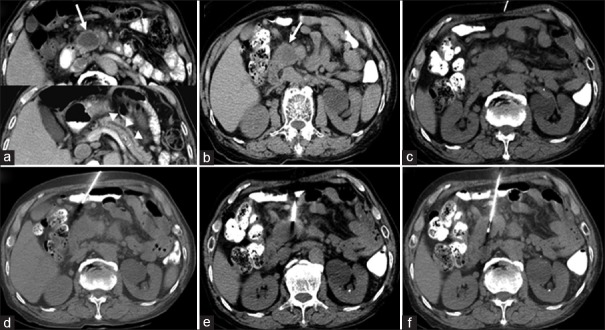
Figure 3.
(a-f) Computed tomography (CT)-guided biopsy images via a fat detour route in an 80-year-old male with a pancreatic head and neck mass. Detour route 1, as shown in Figure 1, was used. The needle insertion angle/path was calculated to avoid going through any organs, and the insertion was one-way. (a) Contrast-enhanced axial CT image revealed a poorly enhancing mass (arrow) within the pancreatic head/neck with a dilated pancreatic duct (arrowheads). (b) Preprocedural CT image showed a hypodense mass within the pancreatic head/neck (arrow). (c) Axial CT image with subcutaneous anesthetic needle inserted. (d-e) Nonenhanced axial CT images showed insertion of the coaxial guiding needle to the level of the pancreatic mass by changing its course to avoid non-target organ penetration (i.e. small intestine) to reach the mass.* (f) The biopsy gun was fired into the pancreatic head/neck mass on the axial CT image. * The needle appears to be traversing the bowel loop because of the partial volume effect of CT images. Briefly, the partial volume effect is the loss of apparent activity in small objects or regions because of the limited resolution of the imaging system. The needle indeed avoided penetrating the bowel because of the detour route, and the route used made it appear as if it traversed the bowel loop

