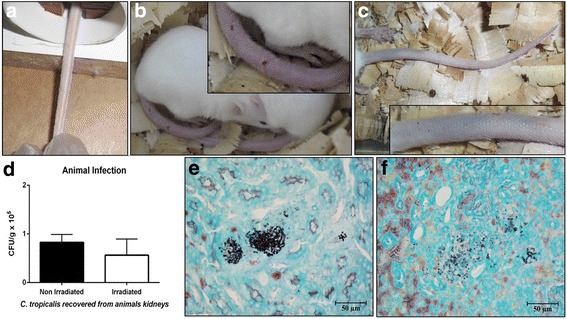
Fig. 2.

Swiss mice (Mus musculus) infected via the caudal vein with clinical isolate of Candida tropicalis after receiving a cumulative dose of 7200 cGy. (a) Tail of no infected animals (before yeast inoculation). The tail of animals infected with irradiated yeasts after 72 h of infection showed a visible inflammatory process, with reddish color, lethargic animals and piloerection (b). Cyanotic tail after 24 h of infection with irradiated yeasts (c). Graph representing yeasts recovered from the kidneys, difference not statistically significant. Data collected from two independent experiments. Standard deviation represented by bars (d). Kidney infected with non-irradiated (e) and irradiated yeasts (f) stained with silver by the Gomori-Groccot technique
