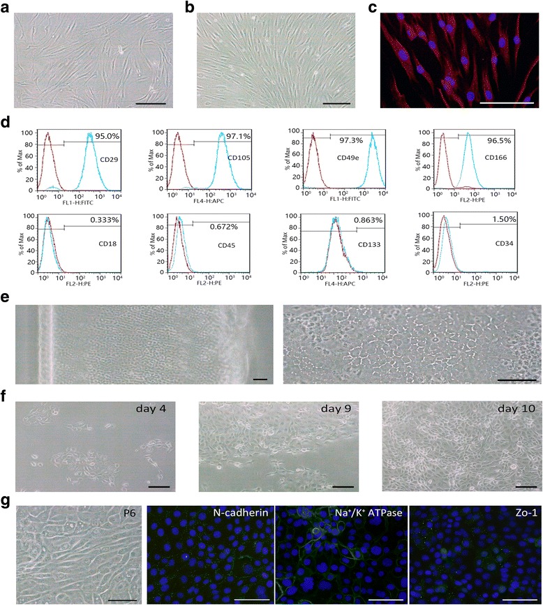
Fig. 1.

Isolation, culture, and characteristics of human orbital adipose-derived stem cells (OASCs) and human corneal endothelial cells (HCECs). a,b OASCs were adherent, spindle-shaped, fibroblast-like cells. c Immunofluorescence staining of vimentin. d Flow cytometric analyses of cell markers in human OASCs (n = 3). Blue lines indicate the OASC group. e Peeled DM layer that contained endothelial cells (left panel) and a magnification micrograph of the DM layer (right panel). f Primary culture of HCECs with OASC-CM (days 4, 9, and 10). g Fibroblastic conversion and expression of CEC relative markers in P6 BM-HCECs. Scale bars = 100 μm
