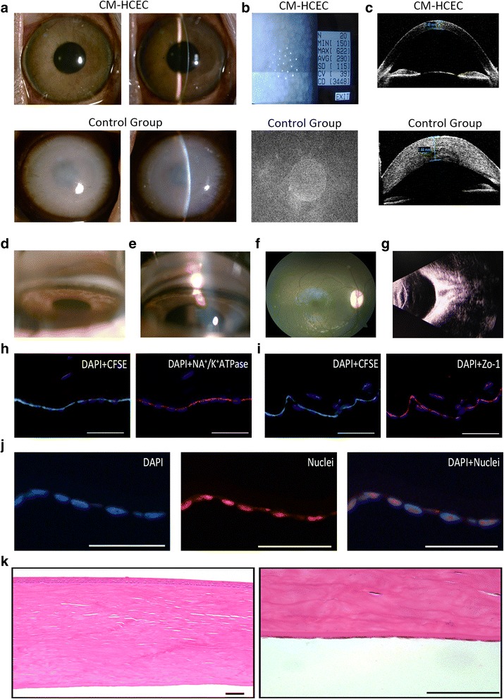
Fig. 8.

Long-term observation after CM-HCEC (P11) injection in the monkey model. a Corneal transparency was examined by slit-lamp in CM-HCEC group and control group. b CECs were detected by noncontact specular microscopy. c Corneal thickness differences in the CM-HCEC group and the control group by OCT. Angle images in the normal group (d) and the CM-HCEC group (e). f Fundus photography in the CM-HCEC group. g B-mode ultrasound in the CM-HCEC group. h Immunofluorescent staining of Na+/K+ ATPase (blue: DAPI, green: CFSE, red: Na+/K+ ATPase). i Immunofluorescent staining of Zo-1 (blue: DAPI, green: CFSE, red: Zo-1). j Immunofluorescent staining of nuclei (blue: DAPI, red: nuclei). k H&E staining of cornea in the CM-HCEC group. Images (a–g) were obtained 10 months after injection. Images (h–k) were obtained 2 months after injection. Scale bars = 100 μm. CFSE carboxyfluorescein succinmidyl ester, CM conditioned medium, HCEC human corneal endothelial cell, Zo-1 zonula occudens-1
