
Fig. 5.
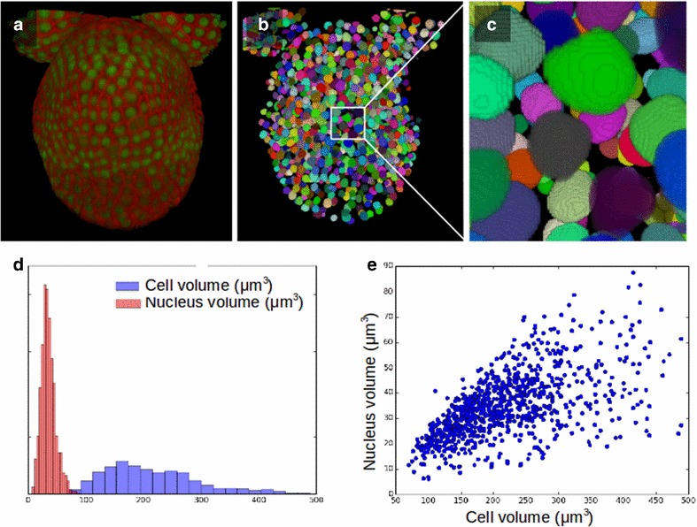
Cellular LSM used for the segmentation of nuclei. a Two-coloured confocal image of an Arabidopsis flower, were cell membrane and nuclei are taken on two distinct channels. b Segmentation of nuclei using level set method (erosion for initialisation , other parameters , , , , type = ‘g’). c Zoom on the segmented nuclei. d Distribution of the volume of nuclei and cells in the tissue. e Nuclear volume versus cell volume, a correlation coefficient of 0.58 was measured