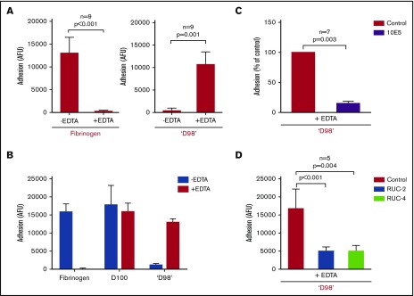
Figure 4.
Paradoxical effect of EDTA in enhancing adhesion of αIIbβ3-HEK cells to fibrinogen fragment ‘D98’. (A) HEK293 cells expressing normal αIIbβ3 (2 × 10³/µL; 50 µL) were added to microtiter wells precoated with fibrinogen (10 µg/mL coating concentration) for 1 hour at 22°C in the absence and presence of EDTA (10 mM) (left). EDTA dramatically inhibited adhesion (n = 9; P < .001). Normal αIIbβ3-HEK cells (2 × 10³/µL; 50 µL) were added to microtiter wells precoated with ‘D98’ (10 µg/mL coating concentration) for 1 hour at 22°C in the absence and presence of EDTA (10 mM) (right). The EDTA dramatically increased adhesion (n = 9; P = .001). (B) Normal αIIbβ3-HEK cells bound to D100 in the absence and presence of EDTA. Conditions as per panel A with D100 coated at 10 µg/mL (n = 3). (C) The mAb 10E5 (20 µg/mL) inhibited EDTA-induced adhesion of αIIbβ3-HEK cells to ‘D98’ (n = 7; P = .003). (D) Small-molecule inhibitors of αIIbβ3 RUC-2 (10 µM) and RUC-4 (5 µM) inhibited the EDTA-induced adhesion of αIIbβ3-HEK to ‘D98’ when added after the EDTA (n = 5; P < .005). Similar results, not shown, were obtained when RUC-2 or RUC-4 were added before EDTA (n = 5; P < .005). Data reported as mean ± SD.

