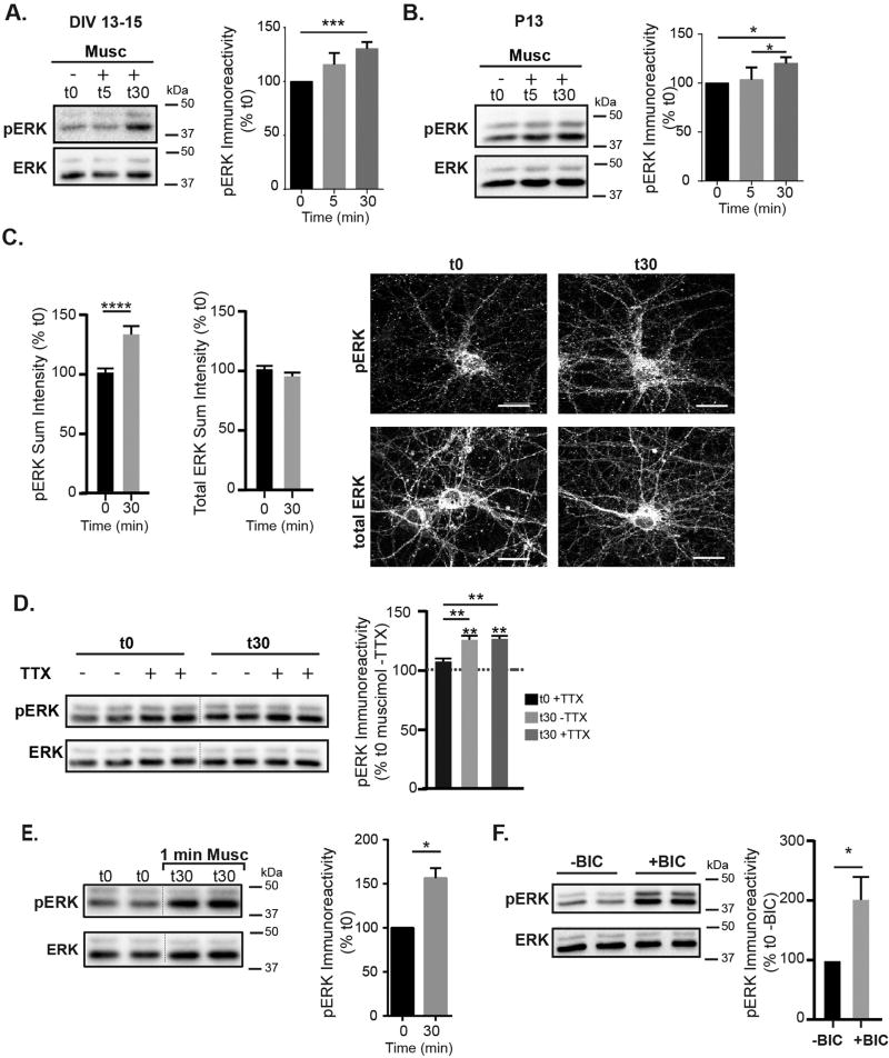
Fig. 3.
GABAAR activity regulates ERK activation. A. Representative immunoblot and analysis of time course for ERK phosphorylation following muscimol treatment at 13–15 DIV. pERK immunoreactivity at t30 was increased compared to t0 (t0 = 100%, t5 = 115 ± 9%, t30 = 130.4 ± 7%, mean ± SEM, n = 6–9 cultures, t30 compared to t0 *p < 0.05, one way ANOVA, Tukey's multiple comparisons test). Total ERK levels were unchanged (t0 = 100%, t5 = 100 ± 12%, t30 = 106 ± 7%, mean ± SEM, n = 6–9 cultures) B. pERK levels were similarly increased in postnatal day 13 rat acute cortical slices: pERK t0 = 100%, t5 = 103 ± 6%, t30 = 120 ± 3%. Total ERK levels were unchanged (t0 = 100%, t5 = 112 ± 8%, t30 = 104 ± 9%. mean ± SEM, n = 4 independent animals, *p < 0.05, one way ANOVA, Tukey's multiple comparisons test. C. Representative immunostaining for pERK and total ERK at t0 and t30 muscimol treatment. Scale bars are 20 µm. Quantification of sum fluorescence intensities, normalized to t0. pERK values increased: t0 = 100 ± 2%, t30 = 121 ± 3 (Mean ± SEM, n = 59–61 neurons, t0 v t30 ****p < 0.0001, t-test). D. TTX does not block the muscimol-dependent pERK increase in DIV 13–15 neurons. Neurons were pre-treated with 1 µM TTX, and then treated for either 0 or 30 min with muscimol. t0 −TTX = 100%, t0 +TTX = 108 ± 3, t30 −TTX = 126 ± 4, t30 + TTX = 126 ± 3 (mean ± SEM, n = 3 cultures, **p < 0.01, two-way ANOVA and post hoc t-tests). E. 1 min muscimol treatment followed by washout produced similar ERK activation to 30 min muscimol treatment: t0 = 100%, t30 (1 min Musc + 29 min washout) = 156.3 ± 11 ((Mean ± SEM, n = 3 cultures,*p < 0.05, unpaired t-test). F. Bicuculline rapidly increases pERK immunoreactivity in DIV 13–15 neurons: t0−BIC = 100.0%, t0+BIC = 201 ± 39 (Mean ± SEM, n = 6–9 cultures, *p < 0.05, unpaired t-test).

