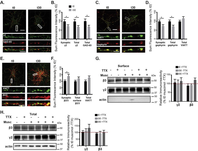
Fig. 4.
Muscimol treatment results in GABAergic synaptic plasticity. A. DIV 13–15 neurons treated with muscimol for 0 or 30 min, fixed, stained for surface γ2, then permeabilized and stained for GAD65. B. Quantification of sum fluorescence intensity of synaptic surface γ2 levels (t0 = 100 ± 10%, t30 = 70 ± 7%), surface total γ2 (t0 = 100 ± 12%, t30 = 67 ± 7%), and total levels of GAD65 (t0 = 100 ± 8%, t30 = 76 ± 8%). n = 40–42 neurons, *p < 0.05, t-test. C. mCherry-gephyrin neurons treated with muscimol for 0 or 30 min, then fixed, permeabilized, and stained for gephyrin and VIATT. D. Muscimol treatment decreased the sum fluorescence intensity of synaptic gephyrin clusters (synaptic fluorescence t0 = 100 ± 8%, t30 = 71 ± 8%, *p < 0.05) and total gephyrin clusters (total fluorescence t0 = 100 ± 8%, t30 = 72 ± 6%,*p < 0.05, n = 41–42 neurons, t-test), with no changes in total VIATT. E. DIV 13–15 neurons treated with muscimol for either 0 or 30 min, fixed, stained for surface β2/3, then permeabilized and stained for VIATT. A,C,E: scale bars 20 µm, and 2 µm on enlargements. F. Muscimol treatment induced an increase in synaptic β2/3 sum fluorescence intensity (t0 = 100.0 ± 8%, t30 = 137.1 ± 13%, *p < 0.05, n = 44–45 neurons, t-test), with no changes in total β2/3 or VIATT sum intensities. G, H. Neurons were surface biotinylated and lysed in RIPA. Immunoblots show that surface and total protein levels of γ2 and β3 subunits, either with or without TTX were unchanged after muscimol (n = 3–6 cultures, ns, two-way ANOVA).

