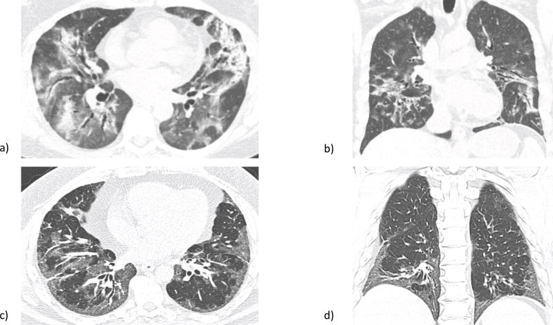
Figure 1.
a) Axial and b) sagittal high resolution computed tomography (HRCT) views of organizing pneumonia (OP), part of the radiographic subdomain of interstitial pneumonia with autoimmune features (IPAF). Notable is airspace consolidation mainly in the periphery. c) Axial and d) sagittal HRCT views of nonspecific interstitial pneumonia (NSIP), which also qualifies under the IPAF radiographic subdomain. Ground glass opacities predominate, particularly in the basilar regions in this patient. Images courtesy of Dr. Jonathan Chung, associate professor of radiology, University of Chicago, Chicago, IL.

