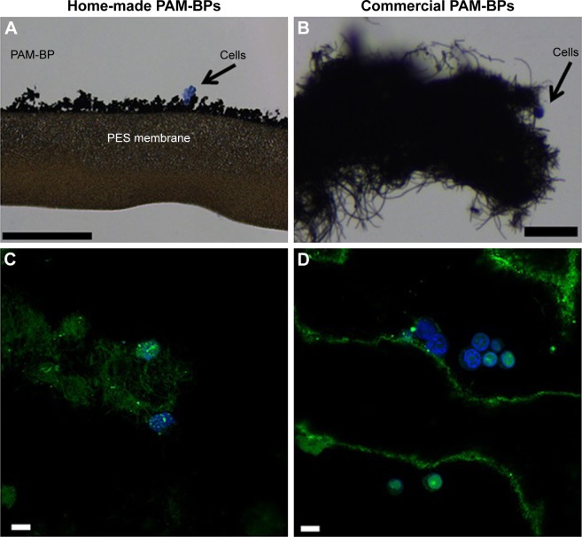
Figure 7.
Cells grown on buckypapers.
Notes: Hematoxylin/eosin staining of cells grown on (A) home-made BP (scale bar =200 μm) and (B) commercial BP (scale bar =25 μm). The commercial PAM-BP incubated with FAM-mir-503 and visualized by confocal microscopy after cell culture (C and D). The green bundles show the intricate network of CNTs forming the BP, whereas the green lines represent the irregular layers of BP. The nuclear staining indicates the presence of healthy cells, whereas the green spots within them indicate that these rigid bidimensional substrates are able to penetrate cells and deliver their nucleic acid cargo. (Scale bar =20 μm.)
Abbreviations: BP, buckypaper; CNTs, carbon nanotubes.

