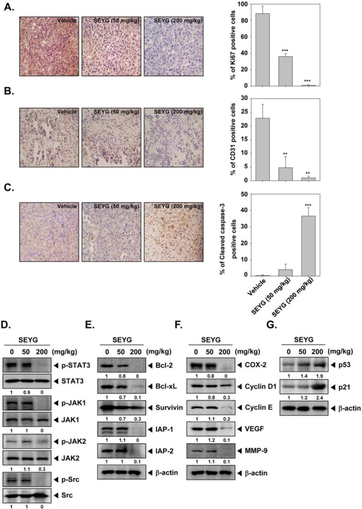
Figure 6.
SEYG exerts the effect against tumor cell proliferation and angiogenesis in prostate cancer. (A) Immunohistochemical analysis of proliferation marker Ki-67+ cell indicates the inhibition of human prostate cancer cells proliferation by SEYG dose-dependent treated groups of animals. Samples from 3 animals in each treatment group were analyzed, and representative data are shown (A, left panel). Quantification of Ki-67 proliferation index as described in “Materials and Methods.” Values are represented as mean ± SD of triplicate (A, right panel). Columns, mean of triplicate; bars, SD. (B) Immunohistochemical analysis of CD31 for microvessel density in prostate tumors indicates the inhibition of angiogenesis by SEYG dose-dependent treated groups of animals. Samples from 3 animals in each treatment group were analyzed, and representative data are shown (B, left panel). Quantification of CD31 angiogenesis index as described in “Materials and Methods.” Values are represented as mean ± SD of triplicate (B, right panel). Columns, mean of triplicate; bars, SD. (C) Immunohistochemical analysis of cleaved caspase-3 in prostate tumors. Samples from 3 animals in each treatment group were analyzed, and representative data are shown (C, left panel). Quantification of cleaved caspase-3 as described in “Materials and Methods.” Values are represented as mean ± SD of triplicate (C, right panel). Columns, mean of triplicate; bars, SD. (D) Western blot analysis showed the inhibition of p-STAT3, p-JAK1, p-JAK2, and p-Src by SEYG in whole cell extracts from animal tissue. The same blots were stripped and reprobed with STAT3, JAK1, JAK2, and Src antibody to verify equal protein loading. (E-G) Equal amounts of lysates were analyzed by Western blot analysis using antibodies against bcl-2, bcl-xL, survivin, IAP-1, IAP-2, COX-2, cyclin D1, cyclin E, VEGF, MMP-9, p53, and p21. β-Actin was used as a loading control. Western blotting samples from 3 mice in each group were analyzed and representative data are shown.

