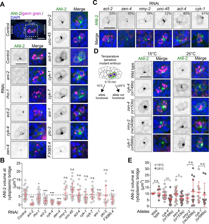
FIGURE 5:
Rho pathway regulators promote ANI-2 accumulation at the stable PGC cytoplasmic bridge. (A, C, D) Confocal images (maximum intensity projections of five to seven consecutive Z planes) of PGCs in 150- to 250-cell-stage embryos, fixed and stained with antibodies against ANI-2 (green), germ granules (magenta), and DAPI (blue). The insets depict PGCs from embryos after RNAi depletion of the genes indicated (A, C) or PGCs from embryos bearing fast-acting, temperature-sensitive alleles of the genes indicated and acquired after continuous growth at permissive temperature (15°C) or after a 5- to 10-min upshift at restrictive temperature (25°C), as depicted in the schematic (D). In C, the number (%) represents the proportion of embryos displaying fragmentation of the ANI-2 focus. Scale bars, 10 μm. (B, E) Quantification of the focus volume of ANI-2 at the PGC cytoplasmic bridge in embryos depleted of the indicated genes by RNAi (B) or in embryos bearing the temperature-sensitive alleles of the indicated genes and maintained at permissive (15°C) or restrictive (25°C) temperature (E). The red bars represent average ± SD. *: p < 0.05; **: p < 0.01; ***: p < 0.001; n.s.: p > 0.05.

