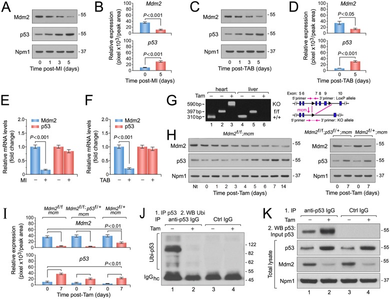
Fig 2. The E3 ubiquitin ligase Mdm2 is indispensable for the negative regulation of p53 protein stability in adult cardiomyocytes in vivo.
Ctrl, control. Hc, heavy chains. IgG, immunoglobulin G. IP, immunoprecipitation. Tam, 4-hydroxytamoxifen. Ubi, ubiquitin. WB, Western blot. Numbers on the right indicate the relative molecular protein weight in kilodalton. (A) Mdm2 and p53 protein levels after ischemic injury (myocardial infarction, MI). Immunoblot analysis of left ventricular extracts (60 ug total protein/lane) of C57BL/6J wild type mice at the indicated time points was performed employing anti-Mdm2 and anti-p53 antibodies as indicated on the left. Animals were 13 weeks old at the time of analysis. For normalization, Western blots were probed with anti-nucleophosmin (Npm1). One representative immunoblot of 3 independent experiments is shown. (B) Protein levels shown in Fig 2A were quantified with ImageJ software. n = 3. (C) Mdm2 and p53 protein levels after acute pressure overload (TAB). Immunoblot analysis of left ventricular extracts (60 ug total protein/lane) of C57BL/6J wild-type mice at the indicated time points was performed employing anti-Mdm2 and anti-p53 antibodies as indicated on the left. Animals were 13 weeks old at the time of analysis. One representative immunoblot of 3 independent experiments is shown. (D) Quantification of protein levels shown in Fig 2C. n = 3. (E) Transcript levels of endogenous Mdm2 and p53 in 10-week-old wild-type mice post-MI as analyzed by RT-qPCR. n = 4. (F) RT-qPCR analysis of endogenous Mdm2 and p53 transcript levels in 10-week-old wild-type mice post-TAB as analyzed by RT-qPCR. n = 4. (G) Heart-specific deletion of Mdm2. Schematic structure of the floxed alleles of Mdm2 (right panel). Genomic PCR results (left panel) of DNA isolated from LV tissue or liver control samples of wild-type (wt), vehicle injected control Mdm2f/f;mcm (-Tam) and Mdm2f/f;mcm mice at 7 days post-Tam (+Tam). Animals were 12 weeks old at the time of analysis. Numbers on the left refer to amplicon sizes in base pairs (bp). lane 1: Mdm2+/+;mcm, LV, -Tam. lane 2: Mdm2f/f;mcm, LV, -Tam. Lane 3: Mdm2f/f;mcm, LV +Tam. Lane 4: Mdm2+/+;mcm, liver, -Tam. lane 5: Mdm2f/f;mcm, liver, -Tam. Lane 6: Mdm2f/f;mcm, liver, +Tam. One representative result of 3 independent experiments is shown. (H) Immunoblot analysis of Mdm2 and p53 levels in left ventricular extracts (60 ug total protein/lane) of Mdm2f/f;mcm (left panel), Mdm2f/+;mcm and Mdm2f/f;p53f/+;mcm mice (right panel) employing specific antibodies as indicated on the left. Animals were 13 weeks old at the time of analysis. One representative immunoblot of 3 independent experiments is shown. (I) Quantification of protein levels of endogenous Mdm2 and p53 in the indicated strains shown in Fig 2H. n = 3. (J and K) Mdm2 regulates p53 protein stability in the adult mouse heart by regulation of its ubiquitin-mediated proteasomal degradation. (J) At 7d post-Tam, Mdm2f/f;mcm mice were intraperitoneally injected with the proteasomal inhibitor MG132 (30 mmol/kg body weight) for 6 hours. Left ventricular lysates were immunoprecipitated (IP) with anti-p53 antibodies or normal rabbit IgG. Ubiquitinated p53 proteins in the immunoprecipitates were identified by immunoblotting with antibodies to ubiquitin. One representative immunoblot of 3 independent experiments is shown. IgG, immunoglobulin G. IP, immunoprecipitation. Ubi, ubiquitin. WB, Western blot. (K) Levels of endogenous Mdm2 and p53 proteins in total left ventricular extracts prepared from Mdm2f/f;mcm mice in the presence and absence of Tam (middle and bottom panels). Samples were subjected to anti-p53 immunoprecipitations and Western blots were probed with anti-p53 antibodies (top panel). The same samples as in Fig 3J were analyzed. The mice were 12 weeks old at the end of the experiment. One representative immunoblot of 3 independent experiments is shown. Fig 2 data are means±s.e.m.

