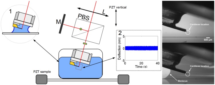
Fig 1. Experimental setup.
A) Schema of the experimental configuration for detecting deflections of a cantilever immersed in a protein solution. A polarizing beam splitter (PBS) divides the input into two beams of crossed polarizations. A mirror (M) reflects the reference beam and provides the overlap of the returned beams. A lens (L) focuses the probe beam at the free end of the cantilever. The reflected beams, probe and reference, interfere after projecting the initial polarizations in the analysis area (see [8] for details). The standard immersion is achieved in a 1.5 ml fluid cell. Inset 1: The meniscus configuration uses a volume smaller than 50 μl. Inset 2: Typical time trace of deflection fluctuations. B) Side view of the cantilever with and without the fluid meniscus.

