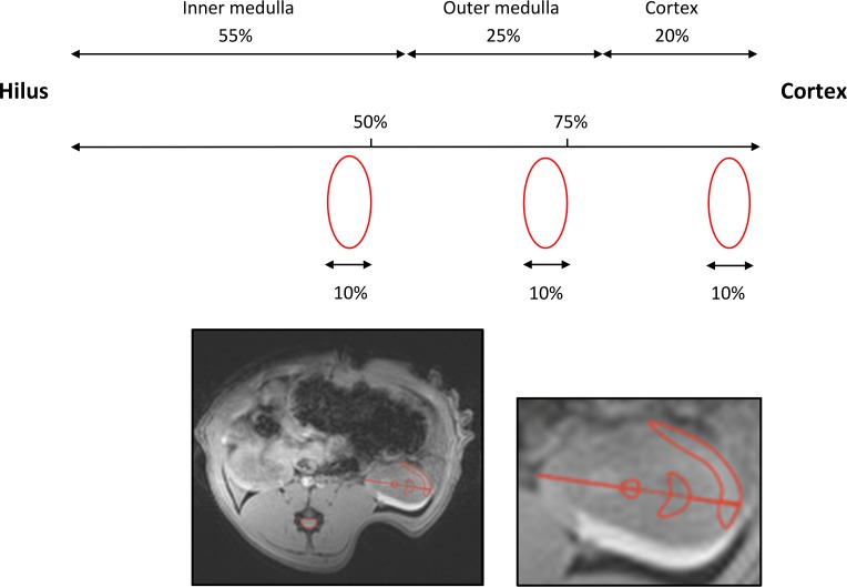
Fig 1. Regions of interest (ROIs).
The size and position of the cortical, outer and inner medullary ROIs were approximated using the morphometrics of the different renal zones from the study by Oudar et al in male Wistar-Kyoto rats (mean body weight 220 g) [31]. The thicknesses of the zones in percent of the total kidney height along the corticopapillary axis were: cortex = 20%, outer medulla = 25% and inner medulla = 55%. First, the central part of the kidney was localized in the axial anatomical T2-weighted image. A vector was drawn starting from cortex ending at the very tip of the hilus/papilla, covering and measuring the whole kidney height. Two marks were placed in the direction cortex to hilus. The first was set to mark the outer medulla and covered 25% of the total kidney height. The second mark, the inner medulla ROI, was put in the centre and covered 50% of the kidney height. The height of each ROI was set to 10% of the total kidney height. The shape of the different zones on a HE-stained cross-section of the kidney were taken into consideration, when drawing the ROIs. The ROIs, we applied, are to some extent similar to the ones proposed by Oostendorp et al [28].

