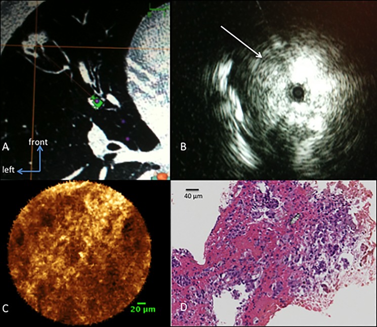
Fig 4. Example of one case with radiologic images of a solitary pulmonary nodule, a respectively r-EBUS image, the pCLE image and corresponding histology.
A: 15 mm solitary pulmonary nodule of the lingula, located at 10 mm of the pleura. B: radial-EBUS signal in this nodule shows a tangential signal on the left part of the image (white arrow). C: pCLE image of this nodule shows a solid pattern on the whole field of view using the 0.6mm CholangioFlex® confocal miniprobe (scale bar: 20 μm). D: H&E staining of the biopsy performed during the procedure shows a pulmonary adenocarcinoma (Magnification x 40; scale bar: 40 μm).

