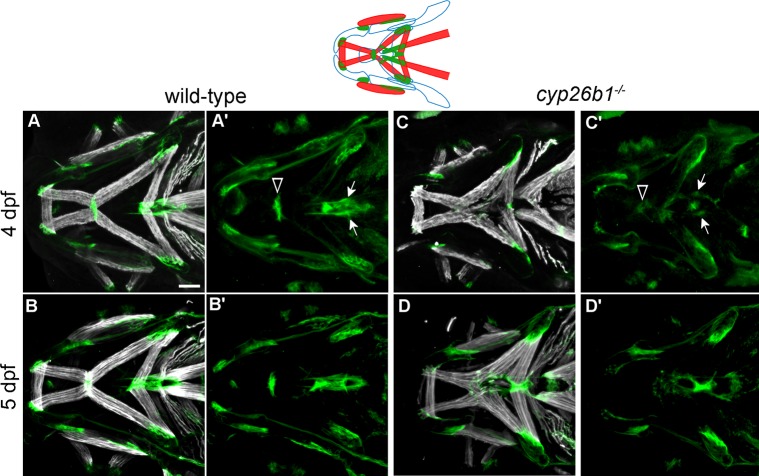
Fig 5. Loss of Cyp26b1 function disrupts cranial tendon differentiation.
(A-B) Tsp4b (green) is enriched at the muscle attachment sites in wild-type zebrafish at 4 and 5 dpf. (C) At 4 dpf, cyp26b1 mutants display Tsp4b at jaw muscle attachments, though weakly at the mandibulohyoid junction (arrowheads in A’,C’) and sternohyoideus tendons (arrows in A’,C’). (D) At 5 dpf, punctate deposits of Tsp4b can be seen at all ectopic points of jaw muscle attachment. All images ventral view, anterior to the left. Scale bar = 50 μm.

