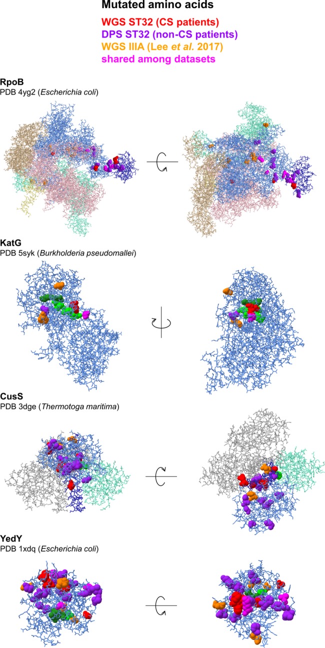
Fig 4. Localization of mutations in proteins under parallel evolution during chronic infection.

Protein chains under parallel evolution are denoted in light blue, their functional domains are denoted in dark blue (DHp in CusS, βi4 in RpoB). Cofactors are denoted as forest green spheres, catalytic and active site amino acids are denoted as lime green spheres. Amino acids homologous to residues affected by mutations during chronic Bcc infection (S10 Table) are denoted as spheres and colored as explained in the legend (for ST32 WGS data, see Fig 3; for ST32 DPS data, see Table 1; for IIIA WGS data, see S9 Table). Visualizations were carried out in Chimera [83].
