
Figure 6.
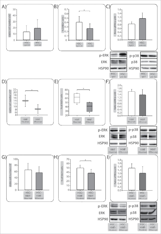
IL-8 is important for HSC-mediated QAP in PDECs while VEGF is a proliferation promoting factor under HMF-coculture. Panc1 cells were indirectly cocultured with (A-C) HSC and either treated with control IgG or an anti-IL-8 antibody. (D-F) Panc1 cells were indirectly cocultured with HMF and either treated with Rituximab as control or Aflibercept to neutralize VEGF. After 6 days, A&D) viable cell numbers and (B) percentage of SABG+ cells or (E) percentage of Ki67+ cells were determined, depicted as mean ± SD or median values with quartiles (Q0,75 as upper, Q0.25 as lower deviation) of 4–5 independent experiments. C+F) Representative Western blots showing expression of phosphorylated Erk (p-Erk) and p38 (p-p38). Hsp90 or Tubulin were used as loading control as appropriate. Data of densitometric analysis of the ratio of p-Erk and p-p38 expression are presented as mean ± SD of 3–4 independent experiments. After 6 d coculture with HSC, coculture of Panc1 cells was prolonged for further 6 d in the presence of HMF (HSC-HMF). (G) viable cell numbers and (H) percentage of Ki67+ cells were determined. I) Representative Western blots showing expression of phosphorylated Erk (p-Erk) and p38 (p-p38). Hsp90 were used as loading control as appropriate. Data of densitometric analysis of the ratio of p-Erk and p-p38 expression are presented as mean ± SD of 3 independent experiments. * = p < 0.05.