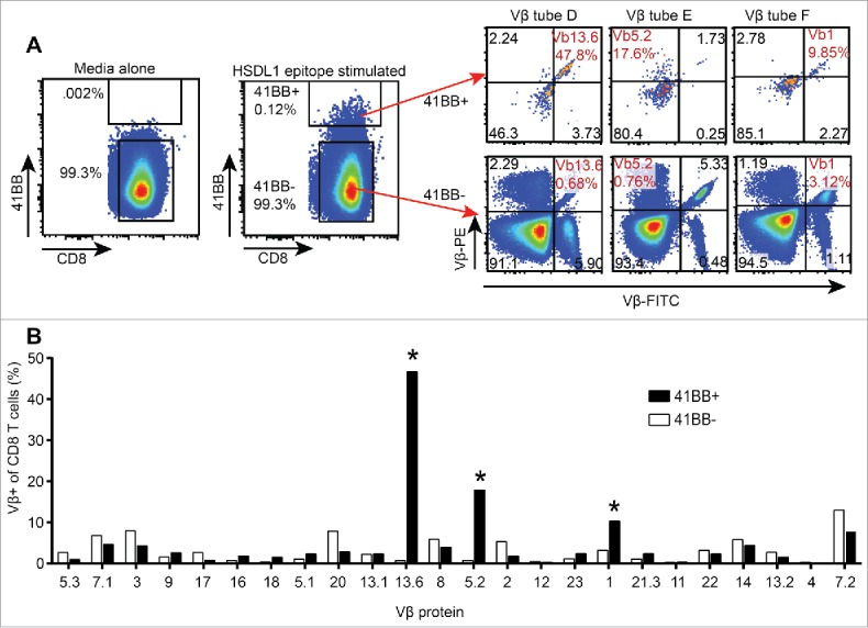
Figure 5.

Vβ spectratyping of expanded TIL from first recurrence ascites. CD8+ TAL were stimulated with the HSDL1L25V peptide epitope or media alone and stained with antibodies to CD8, 4-1BB and each of 8 tubes of the Vβ spectratyping kit. A. HSDL1L25V epitope stimulated CD8+ TAL were gated as 4-1BB+ and 4-1BB−, and the Vβ spectratype plots of 4-1BB+ and 4-1BB− TAL are shown. The Vβ of HSDL1L25V-reactive T cells found in peripheral blood are highlighted in red. B. Bars represent the percentage of all 4-1BB+ (black) or 4-1BB− (white) T cells expressing each Vβ protein. Asterisks show the Vβ of HSDL1L25V-specific T cell lines isolated from peripheral blood.
