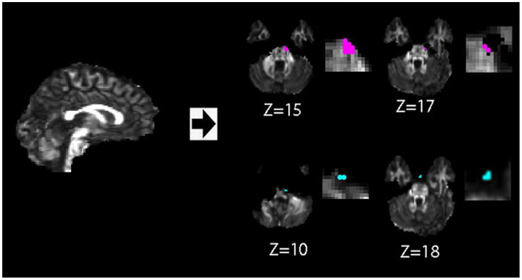
Figure 3.

Location of false positive voxels detected (with zoom in images on the right) using iSPREAD analysis in longitudinal DTI data of one healthy volunteer. False positive of FA (red dots) and MD (blue dots) for iSPREAD are mostly occurred due to image misregistration error or BET residuals, either at tissue boundaries or brain boundaries. Figures are displayed according to radiological convention.
