
Figure 5. Prolonged (15 days) exposure to IFNγ significantly induces immunoproteasome subunit expression and decreases constitutive proteasome subunit expression in astrocytes.
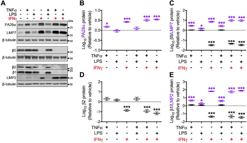
Primary human fetal astrocytes were exposed to TNFα, LPS, and IFNγ (alone or in combination) for 15 days. Media and treatments were replaced every 3 days. (A) Representative Western blot of immunoproteasome (PA28α, LMP7, LMP2) and constitutive proteasome (β5, β2, β1) subunits from a single biological replicate. Quantification of (B) PA28α, (C) LMP7 and β5, (D) β2 and (E) LMP2 and β1 protein expression relative to vehicle after normalization. Data were log transformed and values represent mean ± SEM (n = 4 biological replicates) of the fold change from vehicle (dotted line). Statistical comparisons to vehicle were made by RM-ANOVA with post hoc Holm-Sidak test. *P < 0.05; ***P < 0.001; **** P < 0.0001.