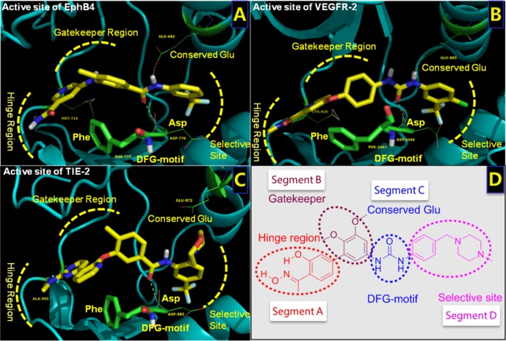
Figure 1. Structures of the active sites of three RTKs bound with co-crystalized inhibitors.
A. Structure of EphB4 complexed with 32W (PDB ID: 4BB4). B. Structure of VEGFR-2 bound with sorafenib (PDB ID: 4ASD). C. Structure of Tie-2 complexed with MR9 (PDB ID: 2P4I). D. Structure and pharmacophore of lead compound (BPS-7). Critical elements of inhibitor interaction are labeled and highlighted.

