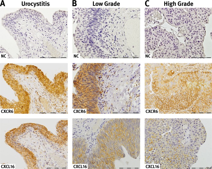
Figure 3. Expression and localization of CXCR6 and CXCL16 in urothelial tissue.
CXCR 6 and CXCL16 immunoreaction in acute urocystitis (A), low grade (B) and high urothelial carcinoma (C). CXCR 6 and CXCL16 are expressed on the surface as well as in the cytosol of epithelial cells. Moreover, in certain areas there is a strong co-localisation of the receptor-ligand pair observable. For detection of negative immunoreactivity (NC) within the same tissue section a consecutive section was stained. The presented slides reveal representative examples of the CXCL16 and CXCR6 immunoreactivity. Magnification, x40; bar 100 μm.

